PRIMARY INTRAMEDULLARY TUMORS OF THE SPINAL CORD AND FILUM TERM IN ALE.
By Johan L.Sloof M.D., James W. Kernohan M.D.,and Collin S. MacCarty '37 M.D. Philadelphia: W. B. Saunders Company, 1964.255 pp. $13.50.
This monograph by two neuropathologists (Drs. Sloof and Kernohan) and the chief neurosurgeon (Dr. MacCarty) of the Mayo Clinic has the special merit of a work focussed on a small but highly important medical field: the tumors which occur and originate within the spinal cord. It carries the authority not only of its eminent authors but of the great volume of case material of the Mayo Clinic gathered over the past 45 years.
The fact that this material is gathered from a single source and that it has been studied and classified with a high degree of uniformity makes this as close to a definitive study of intrinsic cord tumors as we are likely to see. The point becomes obvious when we consider that the 301 primary intramedullary tumors which form the basis of the study were found among over 8000 verified tumors of the nervous system which included 1322 intraspinal tumors.
The first section of the book deals with the difficult problem of determining incidence by comparing the clinical collections of others, the prevalence study done by Kurland on the population of Rochester, Minnesota, and the relative incidence among neurological patients at the Mayo Clinic. There is a detailed analysis of this last material, a brief discussion of clinical symptomatology, physical findings and laboratory diagnosis, followed by a short discussion of neurosurgical treatment.
The main emphasis of the work, however, is on the pathology of the gliomas which account for 273 of the 301 tumors discussed. The classification used is that of Kernohan which is based on the theory that the cells of these tumors represent anaplastic derivatives of normal glial cells. For each of these tumor types there is a detailed description of the histopathology, illustrated with photo micrographs, followed by illustrative case histories and an analysis of the clinical features of each group with separate treatment of the smaller number of tumors of the filum terminale. Nongliomatous tumors are similarly handled.
Thirty-three post-mortem examinations are given individual case study. An appendix gives in tabular form the salient features of each case in the entire group.
For all neurologists — surgical and medical — and, it is hoped, many medical students this will be an essential reference work.
Associate Clinical Professorof Medicine (Neurology)
More From This Issue
-
 Feature
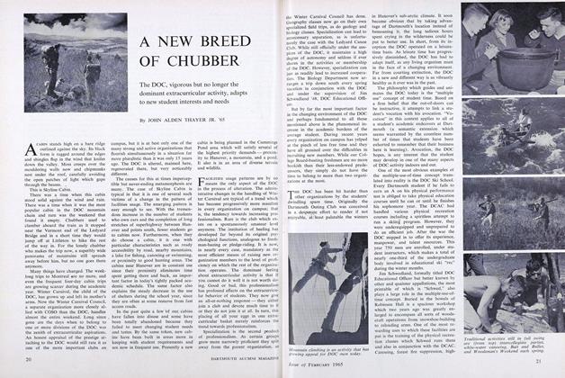
A NEW BREED OF CHUBBER
February 1965 -
 Feature
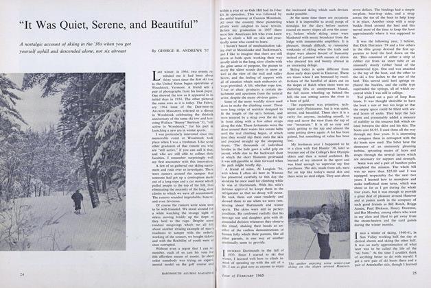
"It Was Quiet, Serene, and Beautiful"
February 1965 -
 Feature
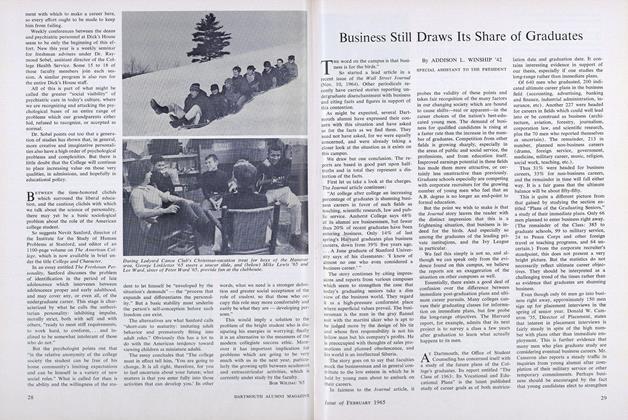
Business Still Draws Its Share of Graduates
February 1965 -
 Feature
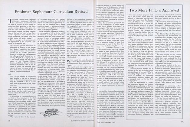
Freshman-Sophomore Curriculum Revised
February 1965 -
 Article
THE UNDERGRADUATE CHAIR
February 1965 -
 Article
Dartmouth's Story Superbly Told
February 1965