Through a Lens Brightly
Discovering an invisible world
IN a small room in Gilman Life Sciences Building is a device that, except for its proximity to a console of switches, dials, and video screens, might be taken for a piece of shiny bar equipment or a futuristic Cuisinart. It is a scanning electron microscope. It cost about $100,000, can magnify an object up to 150,000 times, can produce eerily beautiful and revealing photographs, and according to biologist David Burgess, can be used by just about anybody at the College. Even Coach Joe Yukica, say, if he wanted to examine the laces on a football? Sure, with a little training.
It takes an undergraduate about four or five days to learn to use the microscope competently, says Burgess. Four students, plus faculty from Biology and Geology and the Medical School, have Soloed on the scanner since it became operational six months ago. The procedure involves a complicated fixing process, in which an object - anything up to the size of a golf ball - is dried out, coated with gold dust, and then subjected to a 20,000- volt charge of electrons. The electrons are transmitted into an image on a high-resolution television screen. Besides its capabilities for high magnification and high resolution, an electron microscope differs from a conventional optical microscope in that the object under examination must almost always be dead. The electron charge would take care of that anyway.
At the moment, Burgess is focusing his own research on cell reproduction in sea urchins, while some of the students are studying fresh-water diatoms (minute,highly symmetrical algae) and chick embryos. The electron microscope in Gilman provides startling images of an invisible world, and it has great research potential. "In six months we haven't discovered anything like the double helix," says Burgess, "but we have seen things that haven't been discussed before."
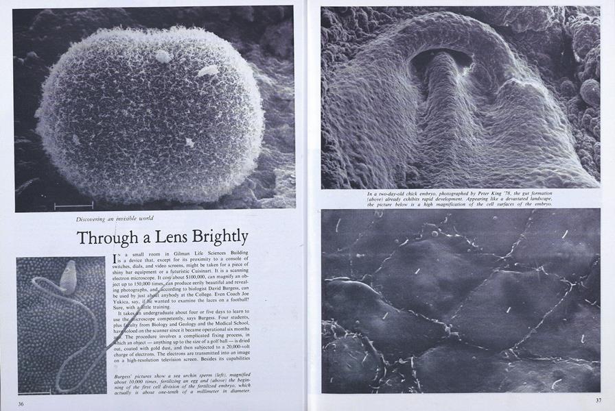
Burgess' pictures show a sea urchin sperm (left), magnifiedabout 10,000 times, fertilizing an egg and (above) the beginningof the first cell division of the fertilized embryo, whichactually is about one-tenth of a millimeter in diameter.
Burgess' pictures show a sea urchin sperm (left), magnifiedabout 10,000 times, fertilizing an egg and (above) the beginningof the first cell division of the fertilized embryo, whichactually is about one-tenth of a millimeter in diameter.
In a two-day-old chick embryo. photographed by Peter King '78, the gut formation(above) already exhibits rapid development. Appearing like a devastated landscape,the picture below is a high magnification of the cell surfaces of the embryo.
In a two-day-old chick embryo. photographed by Peter King '78, the gut formation(above) already exhibits rapid development. Appearing like a devastated landscape,the picture below is a high magnification of the cell surfaces of the embryo.
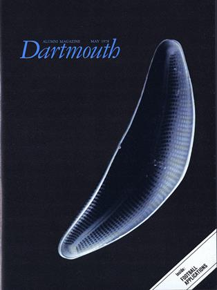
Diatoms take on bizarre - sometimes zany shapesunder the gaze of the electron this page, resembling breadloaves and pasta, was collected from Mud Pond inEnfield, New Hampshire,and photographed by Kenneth Wagner '77 and Professor Nina A lien. They alsotook the highly magnifiedview (right) of the wall of thediatom Epithemia turgida.
The battle for survival goes on at microscopic levels (below): graduate student JeffreyTravis and Professor Robert Allen recorded a marine protozoan at the point of collectingand devouring diatoms with its spattered network of tentacles.
The battle for survival goes on at microscopic levels (below): graduate student JeffreyTravis and Professor Robert Allen recorded a marine protozoan at the point of collectingand devouring diatoms with its spattered network of tentacles.
The battle for survival goes on at microscopic levels (below): graduate student JeffreyTravis and Professor Robert Allen recorded a marine protozoan at the point of collectingand devouring diatoms with its spattered network of tentacles.
More From This Issue
-
 Feature
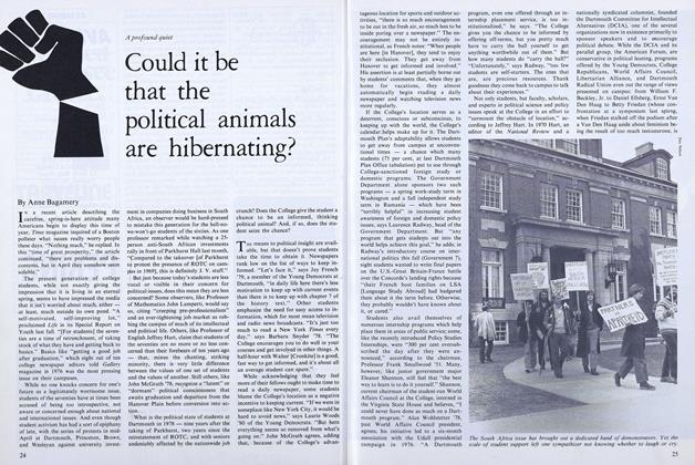
Could it be that the political animals are hibernating?
May 1978 -
 Feature
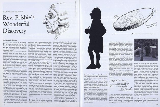
Rev. Frisbie's Wonderful Discovery
May 1978 -
 Feature
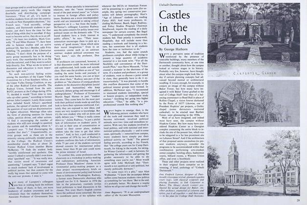
Castles in the Clouds
May 1978 -
 Article
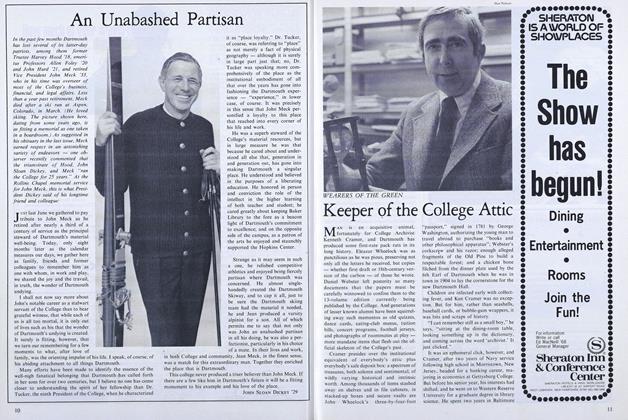
Keeper of the College Attic
May 1978 -
 Article
My Dog Likes It Here
May 1978 -
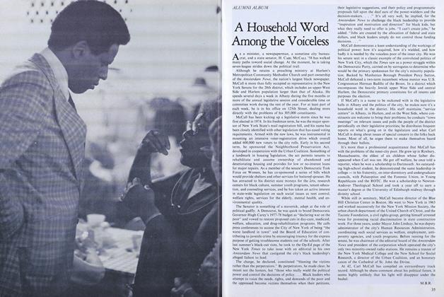 Article
A Household Word Among the Voiceless
May 1978