Dartmouth on the Brain: Green Research and Gray Matter
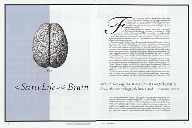
WHICH WAY UP?
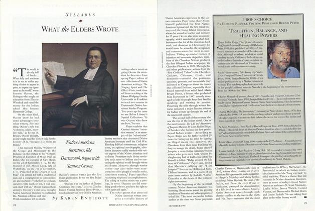
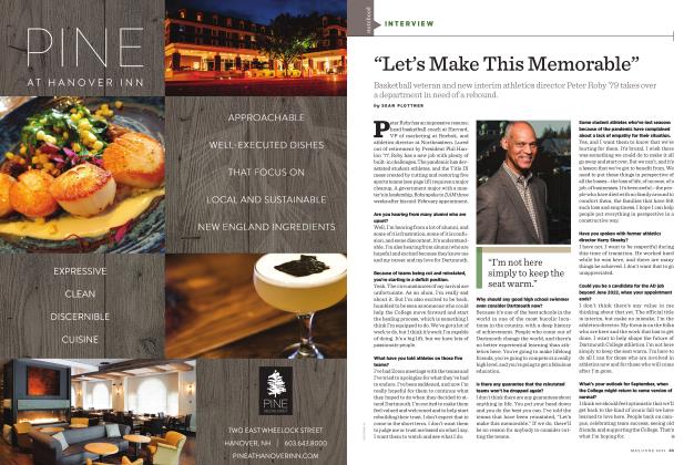
North is north and south is south and never the twain shall meet? Hardly. "It's a very complicated process by which the brain determines left and right, up and down, or north and south," says Dartmouth psychology professor Jeffrey Taube. "Although it feels like second nature, there's a lot going on to tell you your location, your direction, and how to get from A to B."
Taube explores how the mind orients itself spatially. Many people (such as sufferers of strokes, Alzheimer's disease, or motion sickness) experience discomfort or illness when their orienting senses—primarily visual and vestibular (balance)—provide contradictory information. Taube is trying to determine how behavior is correlatec with neuronal activity. Working with the traditional "rat in a maze" scenario, he inserts a tiny wire into specific brain structures and observes which neurons fire when the rat faces different directions. "When they're heading one way, particular neurons go off. When they're going another, a different set fires. So we look to see which sensory cues let the rats know where they are."
Taube also studied the rats on NASA's KC135, an airplane which flies highaltitude parabolas to simulate zero-gravity for 25-second increments. In those spurts he examined how the rats' neurons responded when the animals were placed "upside down" on the roof of their cages. "Frequendy the neurons fired in the opposite way to when the rats were on the floor, suggesing that the rats thought they were turned around 180 degrees," Taube reports. ("I was having the same experience," he says.) So, too, do astronauts, who frequently experience "visual reorientation illusions." When they see crew members working upside down, they suddenly perceive themselves as upside down and the crew members as right-side-up. Potential dangers abound: even turning on a switch depends on spatial orientation.
Taube thinks his rat studies foreshadow what future studies will show about humans. "All of this," Taube says, "leads to a better understanding of how the brain lets us know where we are and where we're heading."
Kevin Goldman '99
ILLNESS AND DENIAL
Working with a wide array of patients with mental disorders, Dartmouth Medical School neuropsychologist Laura Flashman is exploring whether patients are aware of their diseases. Noting that 85 percent of schizophrenics deny having a mental disorder, she says, "You'll ask a patient if they're suffering from this disease they've been diagnosed with, and they'll say 'No.' You can point to concrete evidence, and lots of times they'll even acknowledge some or all of the symptoms, but when you ask if they have the disease, they deny it." The implications are serious. "Patients don't believe they have a disorder, so they don't feel the need to take their medication or continue with their treatment."
To find out why, Flashman is combining neuropsychological testing (general paper-and-pencil tests of memory and other drinking skills) with cognitive activation scanned). She hopes to uncover which areas of the brain recognize—or fail to recognize—that the individual mental disease.
Kevin Goldman '99
BREATHING AND THE BRAIN
What do difficult childbirths, altitude sickness, and near-drownings have in common? Hypoxia sharp reduction of oxygen to the brain.
DMS radiologist Jeffrey Dunn is assessing the brain's ability to adapt to hypoxia. Placing rats, which regulate blood in a similar fashion to humans, in a low-oxygen environment, Dunn and his associates use MRI scans to monitor the metabolic responses and oxygen levels of different regions of the rats' brains. By identifying the critical level of oxygen required for brain function, Dunn hopes to be able to manipulate that level to improve brain survival during hypoxia.
Dunn is also probing oxygen levels in the brain during normal functioning. He stimulates the rats' "whisker barrels" the parts of the brain activated by the whiskers and measures the subsequent change in the brain's levels of oxygen. He hopes that oxygen patterns in the rats will shed light on various responses in human brain oxygenation observed during functional MRI.
Josie Huang '99
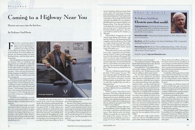
MIND OVER MATTER
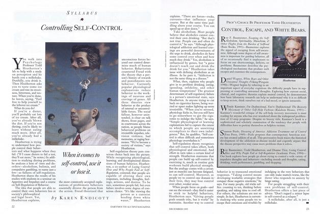
How does the mind form concepts of simple objects? "When you've got ambiguous stimuli, there's something which lets you determine the best way to look at it," says psychology professor Sandeep Prasada. "For example, you could think of something as a table, or you could think of it as hunk of metal. When someone asks you, 'What is this?' there's a bias to say 'It's a table,' rather than 'It's some metal.' But why? What is the mind doing differently when thinking of an entity as an object compared to thinking of .it as stuff?" To find out, Prasada shows subjects different pieces of stuff. Some seems to have a regular form, such as a letter T, while some seems irregular. "When people see the T, it looks like the result of a directed process, so they say it's an object. When they see the one with the irregular shape it doesn't look like it is the result of a directed process, and they say that it's just some wood," Prasada says. But there is power in repetition. "If you then show them identical repetitions of the irregular shape, it doesn't look so random any more, and they say it's an object. Likewise, if you take the irregular-shaped entity and use it to hit a bell, it still seems like just wood—you can hit a bell with anything. But if it fits perfectly into a lock and key mechanism, then it becomes an object. The entity's structure becomes a significant attribute." After Prasada isolates the attributes the mind uses to make these decisions, his next step will be determining which neural mechanisms are involved.
Kevin Goldman '99
THE ANATOMY OF DYSLEXIA
Using an MRI technique that measures the surface area of various regions of the brain, psychiatrist Ronald Green and his colleagues have linked developmental dyslexia to anatomical abnormalities in the brain. Measurements of the planum temporale a region in the right and left temporal lobes of the brain responsible for various aspects of auditory processing (reading is tied to auditory processing) indicate structural differences between non-dyslexics and dyslexics. The plana of dyslexics were larger, and the average difference in size between the left and right planum temporale was much less prominent in dyslexics than in non-dyslexics.
Josie Huang '99
Music ON THE MIND
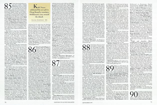
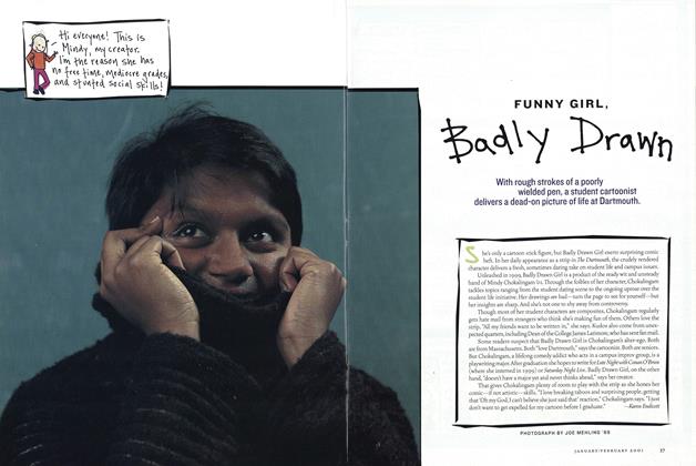
Psychology professor Jamshed Bharucha is using music as a tool for understanding cognition. Working with colleagues Mark Wessinger, Andrew Saykin, graduate student Timothy Justus, and '99s Laura Gibson and Kristin Maczko, Bharucha hypothesizes that the brain's "neural networks" adapt to the musical environment by changing the strengths of the connections between neurons. With fMRI brain scans, the researchers can view which brain regions are activated while a person listens to music. Certain areas of the brain become more active when familiar music is played; the same regions are less active when a person listens to music that, as Bharucha puts it, "violates patterns of one's culture."
Bharucha has also discovered that people with extensive formal musical training exhibit greater activation of the auditory cortex on the left side of the brain than on the right side. People without formal musical training displayed either the opposite or no hemispheric preferences. He offers two possible explanations: either formal musical training results in "greater development of neural circuits in the auditory cortex on the left side of the brain," or certain people who are endowed with the special neural networks will be more receptive to and persevere in their musical training.
Josie Huang '99
THE DEVELOPING BRAIN
Using some 6,000 brain sections painstakingly cut by his own hands, pathologist Miguel Marin-Padilla, a former director of the pediatric autopsy service at the Dartmouth-Hitchcock Medical Center, has been studying genetically abnormal and perinatally-damaged infant brains for clues to normal brain development.
Marin-Padilla's expertise in sectioning the brain led him to correct previous interpretations of the early development of mammalian brains. By sectioning the surface of the brain tangentially rather than at perpendicular angles, he has shown that "Cajal Retzius" cells are horizontal rather than vertical neurons, as previously thought. The cells are among the earliest to appear in the developing mammalian cortex—thus providing a basic framework for subsequent development of all cortical layers.
Marin-Padilla has also been exploring the origin of epilepsy secondary to neonatal brain damage. He hypothesizes that neurons from undamaged cortical regions adjacent to neonatal brain lesions (resulting from premature birth, neonatal asphyxia, and/or circulatory imbalances) undergo a post-injury reorganization. He thinks that this reorganization, not just the original brain damage, plays an important role in causing epilepsy and other neurological disorders in surviving infants.
Josie Huang '99
SMART ROBOTS
Computer science professor Daniela Rus spends her lab time building robots that can perform the same tasks as living creatures. Her "Inchworm" robot can climb doors and fire escapes. In the artificial intelligence class Rus taught last fall, students built "rat robots" programmed with sensors and algorithms that enabled them to navigate mazes and discriminate between edible and poisonous food. Rus is now working on the more complicated tasks of getting several robots to work together to move furniture and designing robots that can metamorphose from one shape into another. "Functionally, it's like building T1000, the robot in Terminator 2 but our robots do no evil," says Rus.
What can robots show cognitive neuroscientists? Whereas biologists explain how living tissue performs a given task, robots provide alternative mechanical models that may result in deeper understandings of organic systems. Robotics also raises the prickly subject of consciousness. Robots can navigate, manipulate, cooperate, and even emulate human emotions, but can a pre-programmed machine ever truly have consciousness? Good question. Maybe too good. Don't look for answers anytime soon, says Rus.
Casey Noga '00
EPILEPSY AND SPLIT BRAINS
Epilepsy is a bane for sufferers, but it's a boon for researchers. Cognitive neuroscience itself grew out of Michael Gazzaniga's study of patients with intractable epilepsy who'd had the corpus callosum, the bundle of nerve fibers connecting the brain hemispheres, surgically severed to reduce the frequency of seizures. Gazzaniga studied patients of Dartmouth-Hitchcock neurosurgeon Donald Wilson, who in the 1970s was performing most of the nation's callosotomies. The 20 patients Wilson operated on and the 100 patients that current Dartmouth-Hitchcock neurosurgeon David Roberts DMS '75 has treated form one of the largest cohorts of split-brain patients in the world.
Dartmouth's epilepsy program is "an intersection of cognitive neuroscience and neurosurgery," says Roberts. "We're looking at the brain everyday. We have an opportunity to learn about the brain—and an obligation to take advantage of the opportunity." The epilepsy program, directed by neurologist Peter Williamson '58, draws from a range of specialties, from neuropsychology to bioengineering.
The collaborations benefit patients as well as scientists. Not that surgery is ever a first resort. It becomes an option only if epileptics fail to respond to anti-seizure medications. For such cases the more researchers learn about which areas of the brain house particular functions memory, language, motor function; to name a few—the more surgeons can fine-tune operations to minimize damage to the mind. And getting into the brain surgically allows neuroscientists to learn more about the how the brain works.
Surgery depends on the type and location of the epilepsy. Callosotomy, for example, is reserved for cases in which all lobes of the brain are involved. Most epilepsy is more localized. The epilepsy team uses tests and imaging techniques to pinpoint the "epicenter of the earthquake," as Dartmouth psychiatrist Howard Hughes calls seizure sites and to assess the impact of surgery. For example, the brain's two hippocampi, common sites for epilepsy, are crucial to memory. "If you don't have at least one working hippocampus, you are unable to remember new information," says neuropsychiatrist Andrew Saykin. Prior to hippocampal surgery, the team injects a fast-acting sedative into the affected side of the brain to temporarily knock it out, then tests the other side of the brain to see whether it can support new memories. A half hour later they repeat the procedure called a Wada test on the other hemisphere for comparison. The results determine the extent of surgery or whether surgery should be avoided altogether.
To pinpoint other seizure sites, Roberts surgically implants a grid of electrodes on the part of the brain expected to produce seizures. Hospitalized and monitored for a week, the patient performs simple tasks—reading words on a computer screen, comparing sounds, counting aloud while the electrodes produce a "cortical map" of brain activity. The procedure is invasive, but helps preserve function when surgery is necessary. The cortical map generated to aid patients has an obvious research payoff as well: it produces working chart of the brain in action.
The search is on for non-invasive access to such information. Saykin predicts, for example, that functional MRI may someday replace the intrusive Wada test. Psychiatrist Jen Schoenfeld, computer scientist Fillia Makedon, and psychology prof Jeffrey Taube are developing a promising technology for assessing spatial memory: a virtual reality probe that will let researchers see how the mine processes information in three dimensions.
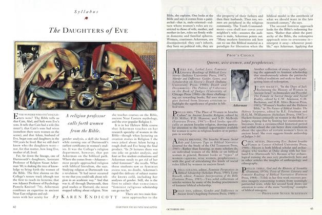
Karen Endicott
WARRING HEMISPHERES
Working from the clinical perspective, DMS neurologist Alexander Reeves has observed a curious, if rare, outcome of splitting the brain via callosotomy to reduce epilepsy: the two sides of a patient's brain can be at odds with one another. Antagonism between hemispheres is usually minor (the dominant hand turns on a faucet, the non-dominant hand turns it off), and most callosotomy patients rapidly develop cooperation between the hemispheres. However, one patient Reeves observed experienced a prolonged and extreme problem. The patient's right and left arms struck each other, and the dominant hemisphere verbally abused the non-dominant arm and leg, as if viewing them as a separate person. Time eventually brought relief. After several years the condition improved.
Kevin Goldman '99
BLINDSIGHT
Cognitive neuroscientist Mark Wessinger collaborates with Robert Fendrich and Michael Gazzaniga on research into the phenomenon known as "blindsight." Normally damage to the visual cortex of the brain results in a loss of vision in the opposite visual field. But in a small number of cases the "blind" can actually see—without knowing they are seeing.
Some researchers attribute blindsight to a secondary visual system in the brain that would supersede a damaged primary one. But in experimental tests, Wessinger, Fendrich, and Gazzaniga found that blind subjects often gave the correct answer about visual stimuli, though they claimed they were only guessing. The subjects were actually somehow processing the visual information unconsciously, the Dartmouth researchers concluded. The blindsight patients were seeing via remnants of undamaged tissue embedded within the damaged area.
According to Wessinger, this visual awareness suggests that consciousness of our surroundings is not relegated to a single area of the brain. The brain may compensate for a deficiency by altering its abilities to process information. That this awareness takes place below the level of consciousness is not unusual, Wessinger says, as our brains must constantly filter out extraneous information about our environment in order to keep our minds focused.
Casey Noga '00
BRAIN GAINS AND LOSSES
As the human brain has evolved, it hasn't only gained function. It has lost some as well. Michael Gazzaniga and colleagues Paul Corballis and Margaret Funnell are investigating the gains and losses achieved through cortical specialization. The researchers are testing their theory by studying the isolated hemispheres of split-brain patients. To test each hemisphere separately, the split-brain patient fixates his eye on a central cross-hair while a stimulus is flashed for 150 milliseconds to the right or to the left. (With this method only one hemisphere can see each stimulus.) The team found that the patient's right hemisphere could accurately judge whether two stimuli were oriented in the same direction or were mirror reversals of each other. The left hemisphere was relatively poor at this and other seemingly simple spatial judgments. These observations led the researchers to theorize that as the left hemisphere became specialized for language, it retained many basic visual skills but lost visual-spatial abilities.
Funnell describes the specialization as an evolutionary tactic that enables the brain to store more functions without requiring additional space. Apparently, as language took over the left hemisphere, it crowded out some of that hemisphere's existing functions. But don't worry about what's going on in your brain. In the normal brain, the researchers explain, loss of a function in one hemisphere should not result in an overall loss of that function, since the two hemispheres communicate via the corpus callosum.
Shirley Lin '02
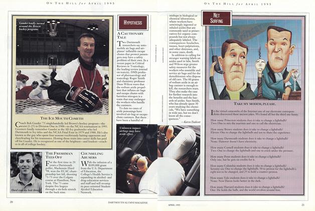
Different neurons fire when rats change directions.
Ceci n'est pas une pipe.
Low oxygen in high placescripples the brain.
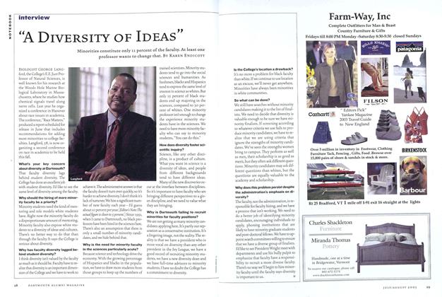
Functional MRI scans show that when a nonmusician listens to music, the right brain is more active than the left (above). Trained musicians experience the opposite effect (below).
"Cajal Retzius" cells form one of the earliest layers of the developing brain.
Brainy robots strut their stuff.
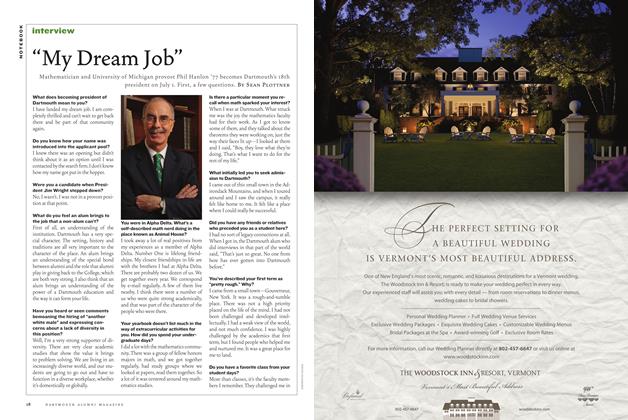
To limit surgical damage to the brain, an electrode grid maps an epileptic's cognitive functioning.
Second century A.D. The Greek physician Galen dissects animal heads. He concludes that intelligence, sensation, and movement originate from the brain. Fourth century Nemesius, a priest, proposes that sensation and imagination are located in the brain's front ventricle, reason in the middle ventricle, and memory in the rear posterior ventricles. Since ventricles are spaces, not tissue, the theory allows Christians to account for human behavior and thought while still believing in a noncorporeal soul. 1363 French surgeon Guy de Chauliac successfully removes a portion of the human brain. The patient live, 1504 Leonardo da Vinci makes wax casts of the ventricles of an ox brain. He believes that the second ventricle is responsible for sensation, not reasoning. 1541 Working from human cadavers, Flemish physician Andreas Vesalius writes the first "modern" anatomy of the brain. The seven volumes contain 15 detailed illustrations of the brain. 1637 Rene Descartes declares"Cogito ergo sum" ("Ithink, therefore I am.").In proposing that mindand body are separate,he tries to please bothchurch and science. Hebelieves the soulresides in the pinealgland near the center ofthe brain. 1780 Luigi Galvani of Bologna debunks Eristratus's "animal spirits" theory of the nervous system. Noticing that frogs legs suspended from copper hooks on an iron balcony were twitching, Galvani shows experimentally that electricity, not spirits, flows through the nervous system. 1810 German scientists Franz Joseph Gail and Johannes Spurzheim co-found phrenology. Hypothesizing that brain functions are localized, they divide the brain into 27 distinct "faculty areas" manifested through bumps on the skull. People feel each other's skulls to analyze personality and intellect. 1822 French physiologist Marie-Jean- Pierre Floerens lops off pieces of pigeons' brains. He finds that the cerebral hemispheres house higher cognitive functions, the cerebellum controls movement, and the medulla controls respiration and other vital functions. 1830 Neurons—nerve cells—are seen for the first time. 1853 Surgeon G. J. Guthrie notes that a person can lose a portion of the back part of the brain without any apparent loss of function. 1848 A dynamite accident blasts an iron rod through the left cheek and top of the head of Vermont railroad worker Phineas Gage. Amazingly, he fives. But with his prefrontal cortex destroyed, Gage's personality changes. The onceplacid and deliberate man now rages uncontrollably and behaves impulsively. 1866 The Journal of Mental Science lists the brain weights of highly intelligent men. The brain of Daniel Webster, class of 1801, weighs in at 53.5 ounces, 2.5 ounces more than the average guy. 1876 Neurologist Carl Wernicke finds a language area in the back of the brain that stores words. 1870sNeurologist John Hughlings Jackson notes that the brain's left hemisphere is dominant for language while the right is better at nonverbal spatial skills. 1884 With ether and chloroform available as anesthetics, brain surgery opens a new window on the brain. 1901 Surgeons Harvey Williams Cushing and Sir Charles Scott Sherrington investigate cerebral localization by probing apes' brains with electrodes and watching their twitching limbs. An aid wields a revolver in case the etherized apes awake. 1903 X-rays are used to pinpoint brain lesions. 1920s During open-brain surgery on conscious patients, neurosurgeon Wilder Penfield stimulates different areas of the cortex. He finds memory areas and maps the language areas. 1929 Psychologist Karl Lashley's mass action theory proposes that since the entire brain is involved in thinking, brain damage will disrupt thinking in direct proportion to the quantity of damage. Lashley tests the theory with maze-running rats. After rats learn a route through a maze, he removes incremental portions of their brains. Lashley reports that the impairment of the rats' memory depends on the volume of tissue removed. Later studies prove him wrong. 1950s Caltech scientists Ronald Myers and Roger Sperry demonstrate brain bilateralism. Experimenting on cats, they sever the corpus callosum that links the brain's hemispheres. The researchers show that information reaching one half of the brain is no longer available to the other half. 1961 Michael Gazzaniga '61 conducts the first cognitive tests on "split-brain" humans. 1980 Positron emission tomography (PET) reveals the structure and biochemical state of brain tissue. The imaging technique is used to diagnose brain tumors, strokes, and mental illness, and to study how the brain functions. 1988 Prozac, a new anti-depressant, demonstrates that the neurotransmitter serotonin affects mood. 1990 The Bush administration kicks off the "Decade of the Brain," to "enhance public awareness of the benefits to be derived from brain research."